If You Give a Mouse a Concussion . . .

LAB PHOTOS BY SIMON BRUTY/THE MMQB
BETHESDA, Md. – One of the most important recent developments in the treatment of brain trauma—and by extension, the future of football—may have been discovered by a clumsy intern.
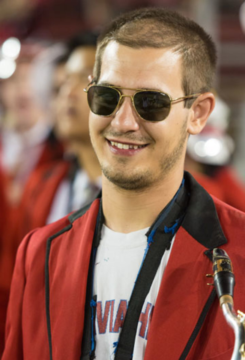
Theo Roth is a St. Louis-born, Alabama-raised Stanford graduate who finagled his way into a National Institutes of Health internship in the summer of 2010, after his senior year of high school. Bored after graduation, he appealed to Dr. Dorian McGavern with a personal email and the recommendation of a mentor of his parents, both doctors. McGavern, new at the NIH’s suburban Maryland health campus, allowed the 18-year-old to sidestep the pool of more than 10,000 college kids gunning for 1,000 spots and took him on. McGavern and his team were using a new research tool, pioneered in 2007 at New York University, which involved shaving down a small portion of a mouse’s skull to shine light into the brain and record its processes. McGavern wanted to study how meningitis affected the brain, but his new intern couldn’t handle the tiny ballpoint saw without concussing the mice and muddling the results.
“He was really bad at performing skull-thinning surgery,” McGavern says of Roth. “Just couldn’t get the hang of it.”
It was—and remains—a difficult thing to accept for a kid who scored 35 out of 36 on his ACTs and 2350 out of 2400 on his SAT. “It’s really hard to do, and they only gave me a week to learn,” Roth says of the procedure. “They have neurosurgeons come in and get it wrong.”
Theo Roth.
Mouse after mouse was concussed, but something valuable did come of the process. Roth and McGavern observed in the subsequent images of the rodents’ brains a flurry of action—leakage from blood vessels lining the skull seeping down and causing brain damage. Towards the end of the summer, the two started talking about what they had seen in the concussed mice. Roth, a former high school wrestler and a Rams fan, connected the dots. “We saw the brain operating in ways no one had ever recorded before, and right around the same time, traumatic brain injury was becoming a hot topic,” Roth says. “People were starting to realize how detrimental it was in the NFL and for guys coming back from Iraq and Afghanistan.”
Roth and McGavern were chatting one morning in the cramped computer room in McGavern’s lab when something clicked. “It was like, wait a minute, we’re actually recreating what happens in a mild traumatic brain injury,” Roth says. “This may actually be very important.”
The wheels started spinning. It became Roth’s personal project to study concussed mice—after all, he was the best in the lab at injuring them. Rather than take his time while sawing down the skull, Roth buzzed through the process in 45 seconds, shaving the bone from a density of one millimeter to 30 microns, about the width of a human hair. The anesthetized mouse was then strapped under a two-photon microscope, a four-foot tall machine that allows for imaging of living tissue. Over the rest of the summer and the summer after that, the bright blue, green and purple images relayed to the computer gave McGavern and his team an outline for the mechanism of damage from minor head trauma. Here’s a thumbnail of what happens:
- A mild brain injury occurs, in this case, when skull is pressed into brain.
- The impact damages blood vessels lining the skull, causing some to burst or leak.
- The body responds, in part, by producing molecules called reactive oxygen species (ROS), which mistake the injury for the intrusion of a foreign body.
- Useful in fighting bacterial infections such as E. coli, the ROS swarm around the injury and cause damage by tearing up the glial limitans, the thin membrane separating the brain from the fluids around it.
- Fluids from the damaged blood vessels leak through the new holes in the membrane and come into contact with brain tissue, destroying it.
McGavern believes this process could play a fundamental role in the development of chronic traumatic encephalopathy (CTE), the disease that has been found in the brains of deceased former football players. CTE has shown up in the autopsied brains of former Bear Dave Duerson and former Charger Junior Seau, both of whom committed suicide. It’s also listed as evidence in the case made against the NFL by thousands of former players who sued over decades of alleged mistreatment of concussions by NFL doctors. CTE and the concussion epidemic are the reason the NFL agreed last year to pay $765 million to those players, plus $30 million to the NIH to fund studies on the biggest problem confronting football at all levels of the game.
But the NFL wasn’t funding this project. This was an NIH undertaking being performed by a researcher whose main interest was viral infection and an intern with shaky hands and a special mind. Roth became obsessed. He went to Palo Alto with mice on his mind. He joined the marching band, and it became his distraction, but as soon as winter and spring breaks began, he was on a plane to the East Coast. “I was much more excited to be doing the research than I was to be taking classes,” he says. “It was a little rough to be studying for a midterm when I was thinking about the next experiment we could do. I was doing work that was brand new, as opposed to learning things from a book that people already learned long ago.”
If You Give a Mouse a Concussion . . .
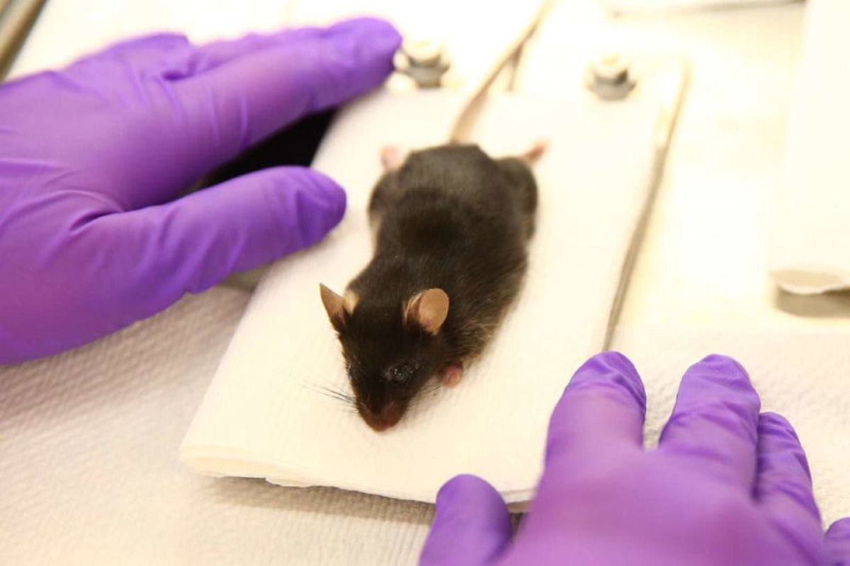
The anaesthetized mouse is prepared for the procedure.
A technician shaves the area of the skull to be studied.
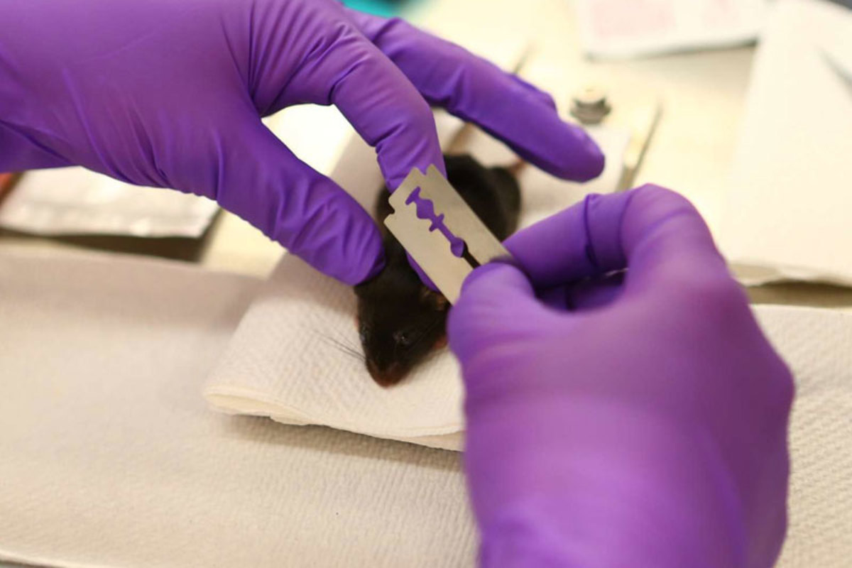
The skull is then shaved down, concussing the mouse.
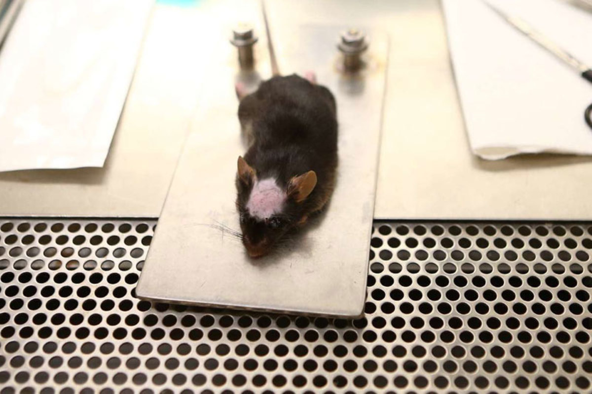
The mouse is secured to a mount.
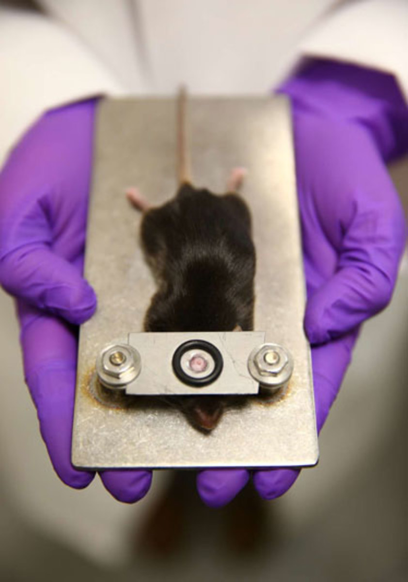
The mount is placed under the microscope.
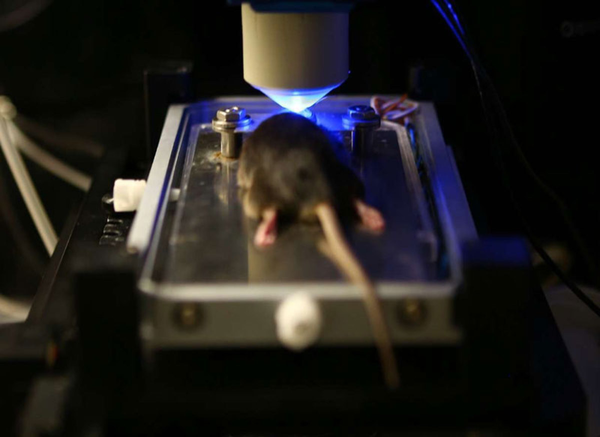
The thinning of the skull allows the microscope to peer directly into the brain.
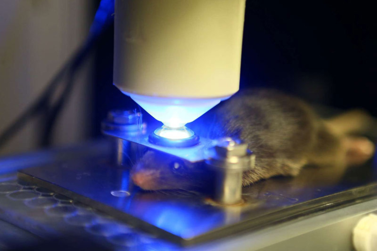
Next, having observed the process of concussion in real time, the researchers brainstormed ways to treat the injury. McGavern remembered that in grad school at the Mayo Clinic, his wife had worked on ROS and ways to block them, for an unrelated study. Her group had used an antioxidant called glutathione. McGavern’s team obtained the readily available organic chemical, and Roth tried it out on mice. He injured the mouse, placed a small quantity of the drug on top of the mouse skull and observed it under the microscope. “You think of the skull as a bone that keeps everything out, but it is a porous filter,” McGavern says.
The morning after their first experiment with glutathione, McGavern arrived at 9 a.m. and found Roth grinning, having slept at the computer. “I saw him, and I already knew that we had it,” McGavern says. “He showed me the result, and the cells look like they were totally naïve. It looks like there’s no injury that’s happened whatsoever. It looks like a normal brain.
Dorian McGavern believes the same treatment his team found reduced brain damage in concussed mice might be effective in people.

“After that Theo lived in the laboratory, 16, 18, 20 hours at a time. He laid his pillow out on the keyboard and would just sleep between experiments. He was a man possessed.”
McGavern reached out to a doctor working a floor below who had been studying concussions in human patients for years. At two local hospitals, concussion patients were given the choice of joining an NIH study for which they were injected with dye and an MRI was taken of the brain. In half of all patients with mild brain injuries, the dye showed up in the brain tissue, which meant the same processes observed in mice were happening in humans.
“You can’t get down to the resolution we can see with the mice, but if you look at the human brain you saw the leakage,” McGavern says.
Roth became the lead researcher on a paper that made national headlines last fall. He reported, among other findings, that passing an antioxidant through the skull immediately after a concussion reduced brain tissue damage by an average of 70%.
(The mice used in the study are able to live normally with their thinned skulls, but typically they’re killed and dissected afterward to further examine the brain tissue. That was no doubt on the mind of dozens of online commenters who threatened McGavern and his team with violence as retribution when The New York Times reported on his research. McGavern has no qualms about using mice in his studies. "When you consider the possible benefit for our species," he says. "It's an easy call.")
Head Trauma in Football

In October The MMQB devoted a week to the most pressing problem facing the game. READ THE SERIES.
Roth was elated. All those spring breaks spent in a lab devoid of daylight, staring at a computer; it meant something now. This was not a cure for brain injury, but it might lead the way to a concussion
treatment
.
“I don’t know if I’ve ever had a person better than him in my lab in 10 years of doing this,” McGavern says. “He’s just brilliant. The amount of information he can collect is ridiculous. And the dedication—think of all the spring break destinations; Cabo, Florida—and instead he was at the lab.”
“An incredible feeling,” Roth says.
Next up for Roth is grad school, after graduation this spring. He applied to 20 graduate programs and got into every one, choosing UC-San Francisco. As for the research, McGavern and Roth’s work spawned two more studies. McGavern’s lab will look into the long-term effects of multiple brain injuries, with and without antioxidant treatment. Within the next several weeks, another researcher at NIH, with McGavern’s help, will study the treatment on pigs, who have skull thicknesses very similar to humans’. Someday, McGavern says, humans could be treated with a dose of antioxidant pressed to the scalp immediately after brain trauma. There’d be no clear telling if it worked, short of the dye MRI and, for football players, a decrease over time in the number of players who suffer loss of brain function, and the degree of that loss.
No doubt Roth would like to get in on subsequent research, though he’s not sure he’s capable of devoting 16 hours at a time to the cause. “I was young and had plenty of energy then,” says Roth, 22. “It feels like I’m getting old.”

